Learn to see microscopically
Rudolf Virchow
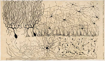
Neuroscience
Neuroscience (or neurobiology) is the scientific study of the nervous system. It is a multidisciplinary branch of biology. Over the years the scope of neuroscience has broadened with new approaches. The study of the nervous system makes use of a wide range of techniques.
In this area –as in many others in biology-, the images analysis is a concept that defines a group of techniques with different objectives that may vary depending on the field of study: Morphometry, Stereology, or 3D reconstruction.
There exist several medical specialties that specifically address the diseases of the nervous system. These specialties also refer to clinical disciplines involving diagnosis and treatment of these diseases. The microscopic observable alterations of some mechanisms of the nervous system are the main focus of research in some neuropathology studies.
Microscopy Images
The images to be studied in these areas are obtained through microscopes. The microscopes are intruments that allow us to see objects that can not be seen by the naked eye. The most common, and also the first to be invented, is the optical microscope. It uses visible light and a system of lenses to magnify images of small subjects.
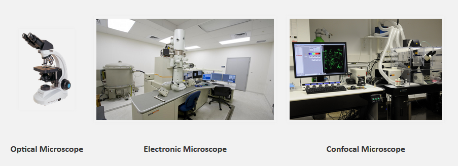
The electron microscopes use a beam of accelerated electrons as a source of illumination. The two major types of electron microscopes are Transmission Electron Microscopes (TEM’s) and Scanning Electron Microscopes (SEM’s). They both have series of electromagnetic and electrostatic lenses to focus a high energy beam of electrons on a sample.
Another interesting microscope is the confocal that uses an optical imaging technique for increasing optical resolution and contrast of a micrograph. In this conventional fluorescense microscope the entire sample is supersatured with light from the illumination source. Due to the conservation of the intensity of light, all parts of the sample will be excited and the fluorescence detected by a photodetector or camera.
M3DSlicer
Microscopy for 3DSlicer
In a first stage of the project, the software application 3DSlicer will be used for the visualization and segmentation of microscopy images. Since this application is composed of numerous modules, extensions and datasets among others, a new extension for the sementation of microsocopy images has been developed.
Natural Neuronal Networks
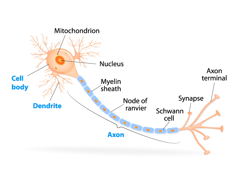
We refer to biological neural network. A series of neurons interconnected that once activated defines a recognizable linear pathway.
The axon terminals are connected via synapses to dendrites on other neurons. and they can be seen as the simplified version of a natural neuron. These nodes connections between artificial networks can be seen as a simplified version of the synapse, they can transmit signals from one to another.
A paradigm of automatic learning and processing inspired by the way the biological nervous system works.
Artificial Neuronal Networks
We refer to computing systems inspired by the biological neural networks. Yes, the ones of the animal brains. Such systems "learn" tasks from examples.
An ANN is a system based on a collection of connected units or nodes. These nodes are called artificial neurons and they can be seen as the simplified version of a natural neuron. These nodes connections between artificial networks can be seen as a simplified version of the synapse, they can transmit signals from one to another.
A paradigm of automatic learning and processing inspired by the way the biological nervous system works.